
CLINICAL EVIDENCE
Seeing what the eye cannot.
DiabSense is being evaluated in real-world clinical environments to explore how thermal imaging can support assessment and monitoring of diabetic and vascular foot complications.
Thermal imaging adds another layer of information
Our clinical work compares thermal patterns with physical examination and established clinical indicators to investigate temperature asymmetry, localized inflammation, reduced peripheral perfusion and tissue changes.
Temperature asymmetry
Compare corresponding regions of the left and right foot.
Localized thermal changes
Visualize areas showing increased or decreased surface temperature.
Clinical correlation
Compare thermal observations with vascular, neurological and physical assessments.
FEATURED CLINICAL CASES
Patterns observed during evaluation
Clinical photographs that may be sensitive are blurred by default and can be revealed by the viewer.
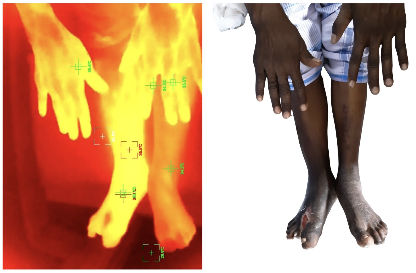
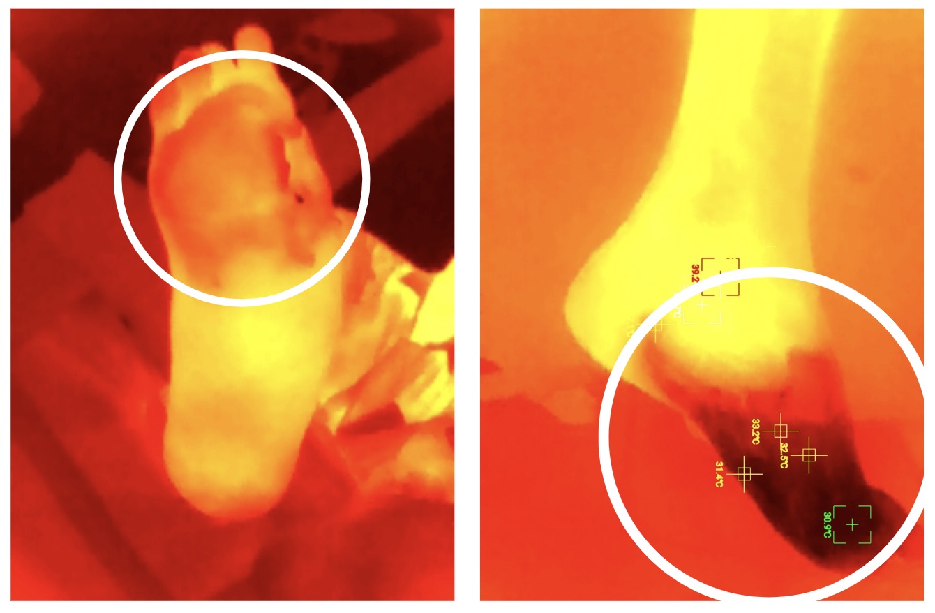
Peripheral arterial disease
60-year-old male · Type 2 diabetes · ABI <0.9
No active ulcer was reported. ABI findings were suggestive of peripheral arterial disease, while thermal imaging showed relatively colder distal/toe regions compared with the lower limbs.
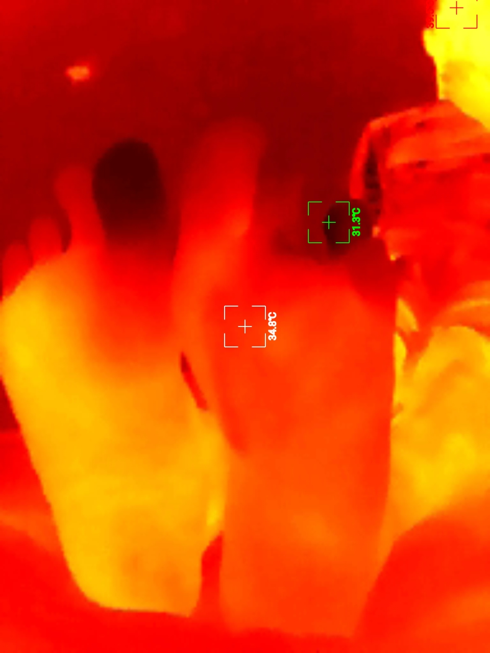
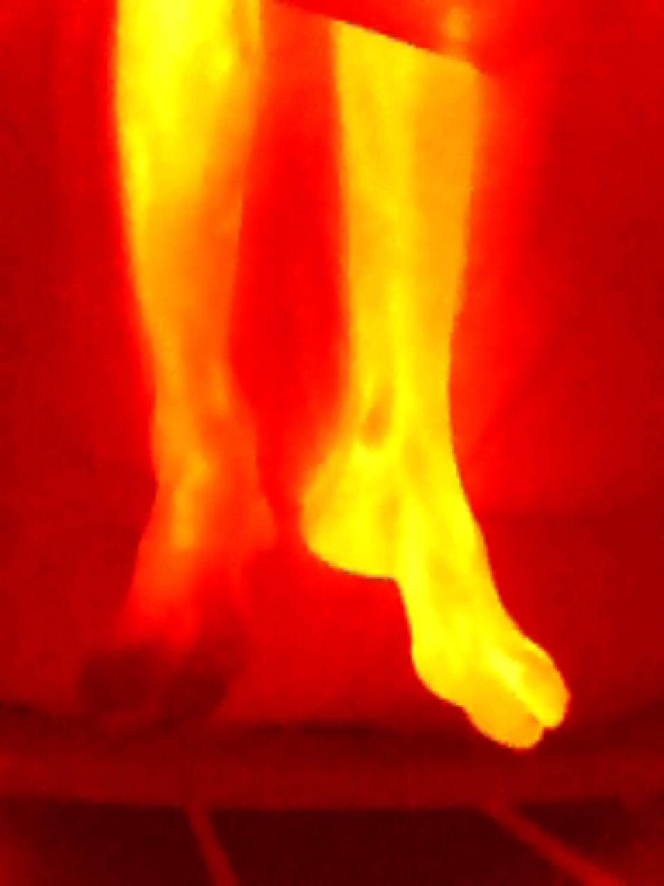
Severe diabetic foot complication
73-year-old male · Type 2 diabetes · Neuropathy · ABI 0.4
Clinical assessment identified an infected deep ulcer, gangrenous toes and significant vascular impairment. Thermal imaging also demonstrated distinct thermal differences around affected tissue.
WHAT WE ARE LEARNING
Clinical context remains essential
Thermal asymmetry
Temperature differences can provide additional information about regional tissue condition.
Distal temperature
Colder distal regions may warrant further vascular assessment of circulation.
Inflammation
Infection and inflammation can increase local temperature and influence interpretation.
Clinical context
Thermal findings should be considered alongside established vascular and neurological assessments.
CLINICAL WORKFLOW
Evidence in context
Clinical interpretation is not based on thermal imaging alone. The repository visuals below help communicate how imaging, patient interaction and clinical review fit together.


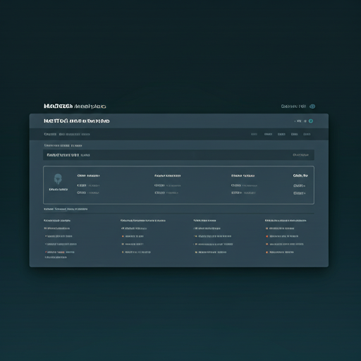
Illustrative interface/workflow visuals; clinical findings on this site remain subject to the research and clinical-use notice above.